由电子科技大学信息与通信工程学院与格拉斯哥大学共同主办,格拉斯哥学院协办的“电子科技大学-格拉斯哥大学学术论坛”,本次邀请到格拉斯哥大学的Sandy Cochran教授及信息与通信工程学院李纯明教授作学术交流。具体安排如下,欢迎感兴趣的师生参加。
一、主 题:
Topic 1:Characteristics of Ultrasound as a Data Source for Medical Imaging
Topic 2:MRI Segmentation and Related Issues Based on Knowledge of Imaging and Anatomy
二、主讲人:
Prof. Sandy Cochran(University of Glasgow)
Prof. Chunming Li(IEEE Fellow, UESTC)
三、时 间:2021年5月19日(周三)下午16:00
四、参加方式:
线上平台:ZOOM Meeting
会议链接:https://uofglasgow.zoom.us/j/92859600520?pwd=cS9qSlRtYWd0TEJyYmRENEhMVmo3Zz09
会议 I D:928 5960 0520
会议密码:424353
五、主持人:Guodong Zhao
六、内容简介:
Prof. Sandy Cochran:Medical imaging with ultrasound is attractive as it is relatively inexpensive, safe and can be applied to provide real-time information at the bedside or in the field. In most cases, the data are generated by an array comprising many elements that transmit ultrasound with controlled timing and receive reflected signals individually. Typically, the signal from each array element is digitised through oversampling at 4x or more with a conversion depth of at least 12 bits. For a typical array of 256 elements operating at a centre frequency of 10 MHz, the resulting radio frequency (RF) data rate, taking into account various aspects of the imaging process, is of the order of 70 MB.s-1, a relatively accessible figure in modern electronics and communications. This is further dramatically reduced through image conversion, realising a video data rate of 14 MB.s-1.
However, two developments in ultrasound that are of increasing interest are its use in intracorporeal applications such as capsule endoscopy and its use in 3-D imaging with 2-D arrays. In the former, the communications channel is very significantly curtailed by attenuation of electromagnetic fields in the human body and, in the latter, the RF data rate may be increased by a factor of beyond an order of magnitude. In this talk, these issues will be explained and the demands of the two developments will be contrasted with the emerging archetype for simple ultrasound systems of a probe connected wirelessly to a mobile phone or tablet. Various pathways to progress will be proposed, without technical detail, and their implications will be considered, particularly for system partitioning.
Prof. Chunming Li:In recent years, data driven methods, represented by deep learning, have been the most popular methods for medical image segmentation. However, data driven methods generally require big training data set with high quality annotation, and their limitations in generalization capability and interpretability often restrict their practical applications. In this talk, I will present our solutions to medical image segmentation and related problems by incorporating knowledge of imaging and anatomy. The talk consists of two parts: 1) From the knowledge of magnetic resonance imaging (MRI), we have the MR image formation model that an image can be decomposed into two multiplicative components with different properties that characterize physical properties of the magnetic fields in MRI devices and the tissues being scanned. To fully exploit the MR image formation model and the properties of the two multiplicative components, we propose a method called scalable local approximation and integration (SLAI) to simultaneously estimate the bias field and segment the brain tissues. The bias field characterizes the inhomogeneity of the B0 and B1 fields in MRI devices, and the estimated bias field can be directly used to perform intensity inhomogeneity correction. 2) From the anatomy of cardiac ventricles, we propose a convexity preserving level set evolution model to recover the endocardium and epicardium of left ventricles from cardiac MRI, such that the obtained endocardial and epicardial contours are convex, which are consistent with the anatomy of the cardiac ventricles. This method is then extended to segmentation of both left and right ventricles by using a novel geometrical structures of cardiac ventricles. Experimental results have demonstrated the advantages of the proposed knowledge based methods for medical image segmentation and bias field estimation in terms of accuracy and robustness.
七、主讲人简介
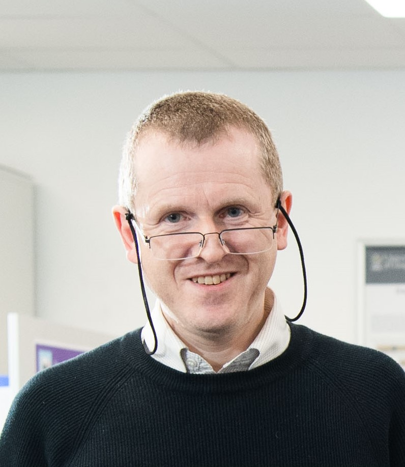
Sandy Cochran (IEEE Member ’00, Senior Member ’19) received the B.Sc. degree in electronics and computing and the M.B.A. and Ph.D degrees from the University of Strathclyde, Glasgow, U.K. He has held posts in engineering, physics, and medicine at several Scottish universities. He is currently a Professor with the James Watt School of Engineering, University of Glasgow, Glasgow, U.K. He has co-authored approaching 500 journals and conference papers and presentations. His research interests are focused on devices and systems for ultrasound in medicine and life sciences. Topics of particular interest at present are new piezoelectric materials and better utilization of existing materials, microscale and miniature devices for new applications, including microultrasound imaging, ultrasound surgery and ultrasound-targeted drug delivery, and ultrasound systems for intraluminal imaging of the gut. Dr. Cochran is a member of the U.K. Institute of Engineering and Technology, and a Fellow of the U.K. Institute of Physics.
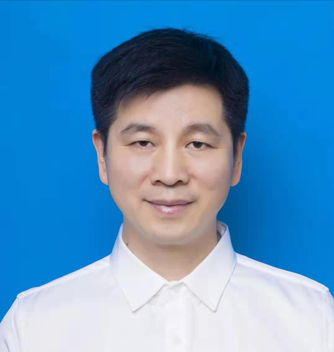
Chunming Li received the B.S. degree in mathematics from Fujian Normal University, Fujian, China, the M.S. degree in mathematics from Fudan University, Shanghai, China, and the Ph.D. degree in electrical engineering from University of Connecticut, Storrs, CT, in 2005. He is currently a full professor in the Department of Information Engineering, University of Electronic Science and Technology of China (UESTC), Chengdu, China. His research interests include the development of innovative algorithms in the fields of image processing, computer vision, and medical imaging. He has published a number of highly cited original articles on image segmentation, level set methods, and related issues in computer vision and medical imaging. He received IEEE Signal Processing Society Best Paper Award in 2013 and 2015 for two of them, both as the first author. He has served as an associate editor or editorial board member for IEEE Trans. Image Processing and Medical Image Analysis. In 2020, he was elevated as an IEEE Fellow for his contributions to computer vision and medical image analysis.
八、主办单位:信息与通信工程学院
协办单位:格拉斯哥学院